Caudal transplantation of ears provides insights into inner ear afferent pathfinding properties
Doctoral student Clayton Gordy and faculty member Professor Hans Straka have investigated how the afferent nerves that sprout from the ectopic ear reach the brain
09.08.2018
The otic placode gives rise to the inner ear in vertebrates. A new study shows that even when it is transplanted to ectopic positions, the nerve cells that grow out of the transplanted ear can form functional connections in the brain.
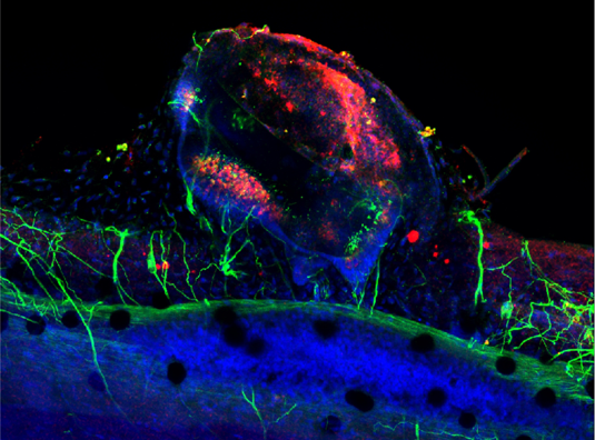
Nerves (green) grow from the inner ear to the spinal cord. Source: Clayton Gordy
In vertebrates, the inner ear develops from the otic placode, a group of cells found in a specific region on the surface of the growing embryo. Studies performed on various vertebrate species have demonstrated that when the placode is transplanted to other sites, it retains the capacity to develop into a normally organized inner ear. In collaboration with Dr. Karen Elliott of the University of Iowa, GSN faculty member Professor Hans Straka and his doctoral student Clayton Gordy have investigated how the afferent nerves that sprout from the ectopic ear reach the brain, and demonstrated the functionality of the connections they make in the brainstem. The new findings appear in the journal Developmental Neurobiology.
Source: LMU News