From Model to Patient - decoding molecular mechanisms of neurodegenerative and psychiatric disease
A complex network of different neuronal populations, their neurotransmitters as well as cerebral endocrine glands and hormones build the basis for major brain and body functions and behaviours. Their disturbance leads to the development of severe human neurological and psychiatric disorders. Using conventional (knock-outs and knock-ins) and conditional mutagenic approaches in mice complemented by a wide array of morphological, histological, molecular/cell biological, biochemical, behavioural and even bioinformatical analyses, we try to understand the physiology, the molecular mechanisms and genetic networks underlying neurogenerative and psychiatric illnesses such as Parkinson's disease and depression.
This combined knowledge may aid in devising new therapeutic strategies for these disorders based on regenerative and/or stem cell-based approaches.
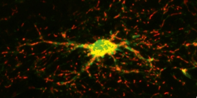 Dopaminergic neurons in the mammalian ventral midbrain
One of the most prominent neuronal populations in the human brain are the neurons located in the ventral midbrain and producing the neurotransmitter dopamine, termed midbrain-dopaminergic (mDA) neurons. The degeneration of these neurons is a hallmark of Parkinson's Disease (PD), characterized by a continuous worsening of motor abilities in the patients suffering from this disease. However, PD patients also experience several non-motor, cognitive and affective symptoms. This is in line with the known and hypothesized functions of dopamine in the human brain: apart from being a key modulator of the motor circuitry in the basal ganglia (the so-called nigrostriatal system), dopamine is also participating in the control and modulation of cognitive (mDA projections to the prefrontal cortex, termed "mesocortical" system) and affective/motivational behaviours (mDA projections to the limbic areas or mesolimbic system).
In order to understand these molecular and functional differences a precise knowledge of the cues controlling the development of these neurons during embryogenesis, which will impart their characteristic projection areas and hence functions is necessary. Furthermore, dissecting the particular function of PD-associated genes in mDA neurons will probably help to understand the pathogenesis of this disease.
We have recently identified a crucial genetic network in the mouse embryo regulating the early establishment of the mDA progenitor domain in the ventral midbrain, and one aspect of the later differentiation of these progenitors into mDA neurons (Prakash et al., 2006) . This network is regulated by a key factor, the secreted glycoprotein Wnt1 that is expressed in the ventral midbrain. Wnt1-signalling maintains the expression of the transcription factor (TF) Otx2, which in turn is required for the repression of the TF Nkx2-2 in this region.
In the absence of Wnt1 and Otx2, serotonergic neurons normally not found in the rostral brain will arise from the ventral midbrain instead of mDA neurons. Therefore, the genetic network comprising these 2 factors (Wnt1 and Otx2) and another secreted glycoprotein, Sonic hedgehog (Shh), is responsible for imparting the mDA neuronal fate to ventral midbrain progenitors (Prakash and Wurst, 2006).
Later in development, Wnt1 probably controls the induction of another key transcription factor, Pitx3. Pitx3 is initially expressed in a subset of mDA neurons but later found in all mDA neurons of the adult mouse brain (Maxwell et al., 2005; Smidt et al., 2004; van den Munckhof et al., 2003).
Notably, the absence of Pitx3 function in the mouse brain leads to a preferential loss of Substantia nigra (SN) mDA neurons (the neurons projecting to the striatum), indicating that Pitx3 has a distinct function in the survival of these two mDA subpopulations. Even more importantly, genetic variations in the human PITX3 gene have very recently been associated with a risk for sporadic PD (Fuchs et al., 2007).
We are therefore focussing on further dissecting the Wnt1-controlled and other signalling pathways in mDA neuron development, and on understanding the physiological function of selected PD-associated genes such as Dj1 (PARK7), Pink1 (PARK6) and Lrrk2 (PARK8) (Farrer, 2006).
in the adult mouse brain by using conventional (knock-outs and knock-ins) and conditional mouse mutagenesis complemented by a wide array of morphological, histological, molecular, cell biological, biochemical and behavioural analyses.
 CRH-dependent signaling mechanisms
Depression is among the most prevalent forms of psychiatric disorders and a major cause of disability with a lifetime prevalence of about 15-20%. According to the World Health Organization unipolar depression is projected to reach second place as leading contributor to the global burden of disease by the year 2020. Depression is considered as a stress-related disorder underscoring the role of stress as a key determinant in disease etiology.
The 41-amino acid neuropeptide corticotropin-releasing hormone (CRH) plays a prominent role in coordinating the neuroendocrine, autonomic, behavioral and immunological responses to various stressful stimuli. Besides its function as the major physiological regulator of hypothalamic-pituitary-adrenocortical (HPA) axis activity, CRH functions as a neurotransmitter/ -modulator, which affects a wide range of behaviors including anxiety-related behavior, arousal, sensory information processing, learning and memory (Figure 4). Dysregulation of the CRH system and accompanying chronically elevated levels of CRH are a hallmark of stress-related and affective disorders, including anxiety disorders and major depression.
In order to dissect CRH-dependent signaling mechanisms special mouse models were developed, which emulate the clinical symptoms of human depression. The observation that patients with psychiatric diseases have elevated levels of CRH in the cerebrospinal fluid, a reduced density of CRH receptors in the frontal cortex, and elevated stress hormone (cortisol) levels in the blood indicates that CRH plays a central role in the development and course of depression and anxiety-related disorders. Changes in the complex interactions between corticotropin-releasing hormone (CRH) and its receptors were also observed.
By genetically ablating the CRH receptor type 1 (CRH-R1) in the mouse brain, it has been shown that CRH regulates anxiety-related behavior in a particular area of the brain, the limbic system - irrespective of its role in the HPA axis (Figure 5). Apart from this, it could also be shown that CRH-R1 function plays an important role in stress-induced alcohol consumption. These results corroborate the significance of CRH-R1 in the development of psychiatric disorders.
The implication of CRH in the pathophysiology of affective disorders has stimulated the interest in a suitable animal model of CRH hyperactivity. In order to study the effects of central CRH hyperdrive, CRH transgenic mouse lines have been established, however, CRH overexpression resulted in elevated ACTH and corticosterone levels accompanied by symptoms of Cushing-like syndrome. To discriminate behavioral effects resulting from central CRH hyperdrive from those resulting from HPA system hyperactivity, we developed a mouse model that permits the overexpression of CRH in a conditional fashion without producing marked neuroendocrine disturbances under basal conditions.
Combining the knock-in of a single copy of the murine Crh cDNA into the ROSA26 (R26) locus with the Cre/loxP system enabled us to overexpress CRH in a spatio-temporally regulated fashion at different dosages (Figure 6). Using nestin-Cre mice to restrict CRH overexpression to the CNS validated this mouse line (CRH-COE-Nes) as an ideal tool to specifically study the effects of increased CRH signaling in behaviorally relevant brain regions and as an extremely useful animal model for screening the efficacy of therapeutically relevant compounds antagonizing CRH-R1. Following this versatile strategy one can achieve any desired pattern of CRH expression depending on the Cre mouse line used. Breeding to different Cre-lines or stereotactic injections of virally based Cre-expression systems may help to further dissect the contribution of CRH-sensitive pathways to the transition from physiological to pathological stress responses that are thought to underlie the etiology of affective and anxiety disorders.
This work is performed in close collaboration with Prof. Dr. F. Holsboer at the MPI of Psychiatry in Munich.
References
Deussing JM, Kuhne C, Putz B, Panhuysen M, Breu J, Stenzel-Poore MP, Holsboer F and Wurst W. 2007. Expression Profiling Identifies the Crh/Crh-R1 System as a Modulator of Neurovascular Gene Activity. J Cereb Blood Flow Metab. 27, 8:1476-95.
Deussing JM. 2006. Animal models of depression. Drug Discovery Today: Dis. Models 3, 375-383.
Schmidt MV, Deussing JM, Oitzl MS, Ohl F, Levine S, Wurst W, Holsber F, Muller MB and de Kloet ER. 2006. Differential disinhibition of the neonatal hypothalamic- pituitary-adrenal axis in brain-specific CRH receptor 1-knockout mice. Eur. J. Neurosci. 24, 2291-2298.
Fuchs J, Mueller JC, Lichtner P, Schulte C, Munz M, Berg D, Wüllner U, Illig T, Sharma M and Gasser T. 2007. The Transcription Factor Pitx3 Is Associated with Sporadic Parkinson's Disease. Neurobiol. Aging.
Farrer MJ. 2006. Genetics of Parkinson Disease: Paradigm Shifts and Future Prospects. Nat Rev Genet. 7, 4:306-318
Lu A, Steiner MA, Whittle N, Vogl AM, Walser SM, Ableitner M, Refojo D, Ekker M, Rubenstein JL, Stalla GK, Singewald N, Holsboer F, Wotjak CT, Wurst W, Deussing JM. 2008 Conditional mouse mutants highlight mechanisms of corticotropin-releasing hormone effects on stress coping behavior. Mol Psychiatry, under revision
Prakash N, Brodski C, Naserke T, Puelles E, Gogoi R, Hall A, Panhuysen M, Echevarria D, Sussel L, Weisenhorn DMV, Martinez S, Arenas E, Simeone A and Wurst W. 2006. A Wnt1-Regulated Genetic Network Controls the Identity and Fate of Midbrain-Dopaminergic Progenitors in Vivo. Development. 133, 1:89-98
Prakash N and Wurst W. 2006. Genetic Networks Controlling the Development of Midbrain Dopaminergic Neurons. J. Physiol. (Lond). 575, 2:403-410
Maxwell SL, Ho HY, Kuehner E, Zhao S and Li M. 2005. Pitx3 Regulates Tyrosine Hydroxylase Expression in the Substantia Nigra and Identifies a Subgroup of Mesencephalic Dopaminergic Progenitor Neurons During Mouse Development. Dev. Biol. 282, 2:467-79
Muller MB, Zimmermann S, Sillaber I, Hagemeyer TP, Deussing JM, Timpl P, Kormann MS, Droste SK, Kuhn R, Reul JM, Holsboer F and Wurst W. 2003. Limbic Corticotropin-Releasing Hormone Receptor 1 Mediates Anxiety-Related Behavior and Hormonal Adaptation to Stress. Nat Neurosci. 6, 10:1100-7
van den Munckhof P, Luk KC, Ste-Marie L, Montgomery J, Blanchet PJ, Sadikot AF and Drouin J. 2003. Pitx3 Is Required for Motor Activity and for Survival of a Subset of Midbrain Dopaminergic Neurons. Development. 130, 11:2535-2542
Sillaber I, Rammes G, Zimmermann S, Mahal B, Zieglgänsberger W, Wurst W, Holsboer F, Spanagel R. 2002 Enhanced and delayed stress-induced alcohol drinking in mice lacking functional CRH1 receptors. Science 296:931-p33
Smidt MP, Smits SM, Bouwmeester H, Hamers FP, van der Linden AJ, Hellemons AJ, Graw J and Burbach JP. 2004. Early Developmental Failure of Substantia Nigra Dopamine Neurons in Mice Lacking the Homeodomain Gene Pitx3. Development. 131, 5:1145-1155
Timpl P, Spanagel R, Sillaber I, Kresse A, Reul JM, Stalla GK, Blanquet V, Steckler T, Holsboer F and Wurst W. 1998. Impaired Stress Response and Reduced Anxiety in Mice Lacking a Functional Corticotropin-Releasing Hormone Receptor 1. Nat Genet. 19, 2:162-6